Abstract
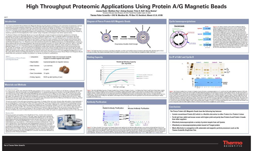
Speed and sample purity are key issues for proteomics researchers. Sample and data-processing throughputs are current bottlenecks in proteomic analysis. Magnetic separations can provide greater consistency and automation of proteomic sample preparation. We report here the development of a novel 1µm protein A/G magnetic bead that enables fast and convenient isolation of proteins via immunoenrichment. Thermo Scientific Pierce Protein A/G Magnetic Beads use a recombinant protein A/G that combines the IgG binding domains of both protein A and protein G. Protein A/G contains four Fc-binding domains from protein A and two from protein G, making it a convenient tool for capture of immunoglobulins from a variety of species and isotypes. Recombinant protein A/G was covalently coupled to 1 μm, blocked double-shelled magnetic particles with a polymeric core. The beads had a binding capacity of >60 μg rabbit IgG per mg bead along with low non-specific binding. Using a Thermo Scientific KingFisher Flex Magnetic Particle Processor, antibodies were purified from serum or ascites with high yield using the Pierce Protein A/G Magnetic Beads. The beads were also used to immunoprecipitate and co-immunoprecipitate multiple proteins significant to different phases of the cell cycle, including cyclin B, Cdk1, cyclin E, and cyclin D. U2OS and HeLa cell lines were first synchronized in serum starved media. The media was replaced with enriched media and cells were harvested at 4 hours (beginning of G1 phase), 6 hours (end of G1 phase), and 18 hours (end of G2 phase). Proteins were effectively isolated from the cell lysates using Pierce Protein A/G Magnetic Beads and identified by Western blot. In addition, these beads were successfully used in a chromatin-immunoprecipitation (ChIP) assay to measure binding of RNA Polymerase II to the proximal myc promoter in A431 lung carcinoma cells treated with and without EGF. In Conclusion, the results indicate that Pierce Protein A/G Magnetic Beads can effectively isolate protein targets in a variety of proteomic applications.
Citation Format: {Authors}. {Abstract title} [abstract]. In: Proceedings of the 102nd Annual Meeting of the American Association for Cancer Research; 2011 Apr 2-6; Orlando, FL. Philadelphia (PA): AACR; Cancer Res 2011;71(8 Suppl):Abstract nr 4877. doi:10.1158/1538-7445.AM2011-4877
- ©2011 American Association for Cancer Research
